From brain signals to computer commands, part 1: acquiring signals
An introduction to signal acquisition with invasive neural recording techniques
Signal acquisition is arguably the most important step in building devices that interact with the brain
In this post, I will discuss invasive recording techniques and the acquired brain signals that are used in invasive brain-computer interfaces (BCI). The cons and pros of invasive BCIs. And finally a glimpse into the future.
When building brain-computer interfaces and neurotechnology, the very first step is to acquire a signal from the brain. As such, brain signal acquisition is one of the most important components of a BCI. Let’s begin by discussing both invasive and non-invasive techniques for signal acquisition that are in use today. The drawbacks and benefits of the respective methods. Afterwards diving into the current status, challenges, and future developments of the invasive methods.
We will begin by once again taking a look at the components of a BCI:

As the diagram above shows, there are generally five components in building a BCI. These are signal acquisition, signal pre-processing, feature extraction, classification, and finally the application interface. In this post I focus on the first step: Signal acquisition for invasive BCIs.
Although before diving into signal acquisition, let’s do a quick recap of the general principles of BCIs.
What’s a brain-computer interface?
A BCI is a device that acquires brain signals in order to analyze and translate them into commands that that carries out a desired action.
BCIs stand apart of other brain-controlled systems when interacting with the external environment, such that it does not require any neuromuscular activity. It converts thoughts into action with no means of muscular control.
That’s amazing! It’s basically the technological equivalent of telepathy and psychic powers 🔮
I can’t stress this enough, it’s a huge deal! Specially for people suffering from neuromuscular degenerative diseases or those who have lost a limb. BCIs allow for the replacement of disrupted neuromuscular pathways of humans, restoring the motor functions of disabled people, or turning simple prosthetics into wearable robotic devices. BCIs are the beginning of the end of disorders such as ALS, stroke-related disabilities, spinal cord injuries, cerebral palsy, muscular dystrophies, multiple sclerosis, and disabilities due to loss of limb. (just to name a few)
…and just imagine the potential in healthy humans!

Electrical activity in the brain
Before we can dive into signal acquisition from the brain, we have to understand how the brain communicates within its modules and layers. The primary takeaway from this section should be: the brain speaks two languages: one metabolic, one electric. (= it’s electrochemical)
(leave a comment if you’d like to see an article on the topic of modules and layerings of the brain & consciousness)
Today I’m feeling sparky, so I’m focusing on the electric signals ⚡️

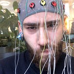
The fact that there’s electrical activity in the brain was first reported in several studies in the late 18th century. However it was not before 1924, Hans Berger (1873 – 1941), a German psychiatrist, became the first to study the electrical signals in the brain. Five years later, he could attribute himself as the inventor of electroencephalography (EEG), which is the phenomenon of measuring the electrical activity of the brain through the scalp. Since then EEG signals have been widely studied to identify the characteristics of brain disorders.
This gave us a tool to clinically categorize brain disorders with a new dimension, which inspired scientists to develop new methods of reading the electrical activity of the brain. These include other non-invasive methods such as magnetoencephalogram (MEG). Likewise semi-invasive and invasive methods have also been developed. The reason for developing more invasive methods are - among other things - reduction of signal-to-noise ratio and gaining a higher spatial resolution.
Electrocorticogram (ECoG) is basically like an EEG, although instead of on the scalp, the electrodes used for measurement are placed directly on top of the brain. Therefore, we define them as a semi-invasive technique.
Intra-cortical, or in daily talk microelectrodes, such as the Utah arrays are a fully invasive recording technique, wherea bunch of small needles are implanted into the surface of the brain.
Due to the invasive characteristics of these techniques, it naturally requires proportionally incapacitating clinical problems, before being taken into use. (in humans at least 🐭🪤)

The main difference between measuring metabolic and electrical activity, is that while non-invasive electrical measuring techniques offer a high temporal resolution, they suffer from both high level of noise and furthermore they are computationally complicated to handle since all sensors record almost the same signal. (known as the superposition of all brain activity). Therefore they must be disentangled (e.g. statistically) for optimal performance.
Adversely, metabolic signals offer a high spatial resolution in multiple dimensions, but require a large amount of resources (big expensive scanners, e.g. MRI machines), and have a low temporal resolution. Further, the metabolic signals require high levels complexity in computation as well.
One solution to both challenges are measuring the electrical activity invasively!


Invasive recording
It should come as no surprise: in order to implant an electrode array into the brain, invasive recording techniques involve a surgical procedure. The electrode penetrates through the brain tissue and is capable of recording neuronal level potential changes of the brain.
Using invasive recordings is far superior to existing non-invasive BCI alternatives, resulting in higher spatial and temporal resolutions, lower signal-to-noise ratio, and increased bandwidth.
Naturally, the majority of research in invasive techniques is done with the help of animal studies. Throughout the years, studies have successfully been completed with animals controlling robotic limbs in self-feeding tasks, including orientation and grasping of objects.


With brain penetrating electrode arrays the signal are either read as spiking activity of single neurons with potentials in the range of 5–500 µV and frequencies of 0.1–7 kHz or as Local Field Potentials (LFPs) with electrical potentials < 1 mV and frequencies of < 200 Hz.
Spiking activity from single neurons is measured in the extracellular space, and is recorded by using a high sampling rate. These signals are typically characterised by being either single unit activity (SUA, being sourced from a single neuron) or multiunit activity (MUA, sourced from multiple neurons). With the current technological limitations, continuous SUA measurements on the same neuron has proven quite difficult. Chronically implanted microelectrode arrays are able to record continuously for years on and on, but the neuron population they record changes constantly.
LFPs are the result of the oscillations of the membrane potentials of neuron populations close to the recording electrode. As positive and negative membrane potentials cancel each other out, the synchronized activity of the respective pool of neurons can be detected.

In short, think of LFPs as the summed activity of synchronous electrical activity of ‘local’ brain tissue. ‘Local’ tissue meaning, cells near the respective electrodes.
Almost all current fully invasive BCIs use either one or more Utah arrays. Some are in development that aims to provide neuronal feedback to the patients, but none of the devices have, as yet, been approved for commercialization or use outside of animal and clinical studies.
Although, the potential of giving neuronal feedback to the linked brain, is yet another benefit that invasive BCI systems will have over its semi- and non-invasive counterparts, allowing for somatosensory feedback.
The limitations, besides being invasive and requiring surgery, include that the recording microelectrode usually only occupies a relatively small area of the brain cortex. A restriction that highly limits insights on the dynamical processes on an intercortical level, allowing us only to observe small neural populations.
Further, it can never be garranteed that the tissue won’t begin to induce an immune reaction resulting in inflammation and gliosis. At best potentially resulting in the decrease of neurons close to the recording electrodes.
Behold the future
Many have come to know Elon Musks Neuralink venture. While Musk has yet long way in achieving the same level of disruption in neurotechnology, as he has in electrical vehicles and space rockets, his company has shown some incremental improvements in the development of invasive recording techniques. Specifically two developments stand out:

In 2019 Neuralink demonstrated a ‘sewing machine’ that can implant recording electrodes with minimal tissue damage.
Last August (2020) Musk demonstrated a coin-sized chip implanted in a pig, showing real time recordings. Also presenting a pig, which had the chip implanted and removed once again, showing reversibility of the tool.
In July the Texas-based company Paradromics presented a 65.000 channel implant (in comparison, the BlackRock Utah Array has 100 channels).
All the while, a third company Synchron is experimenting with endovascular stents, foregoing the need of opening the skull surgically.
Read more about the recent developments in Emily Mullin’s beautifully written medium post for Future Humans from December 2020.
Personally, I believe that nano-lab-on-chip particles delivered by vascular means, will define the long-term future of (minimally)-invasive BCIs.
A personal message:
Help me write better and more posts on neurotechnology by giving feedback or requesting topics to cover.
The next posts in the series will be on signal acquisition in non-invasive (primarily EEG) based BCIs, followed by a post on signal pre-processing.